%20Gradient%20%E2%80%94%20light%20to%20dark.png)
X-ray imaging is one of the most powerful tools for characterizing battery cells, but X-rays are also known to damage certain materials at high doses. That raises a natural question for anyone scanning batteries: does the imaging itself harm the cell? Despite how widely CT is used in battery research and quality control, we were unaware of previous reports that systematically studied whether lab-scale X-ray exposure affects battery performance and lifetime. We decided to find out.
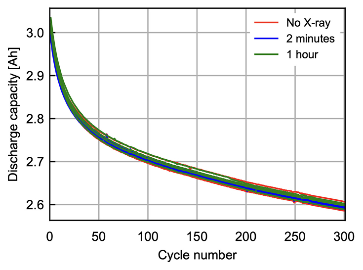
Capacity retention curves for cells exposed to 0, 2, and 60 minutes of X-ray exposure, showing no statistically significant difference across groups. Reproduced from Figure 2 of Min et al.
The result is Glimpse’s latest publication, led by our former summer intern Jinhong Min. The paper, entitled “Lab-Scale X-Ray Exposure Has No Measurable Impact on Lithium-Ion Battery Performance and Lifetime,” was published in Batteries and can be read here.
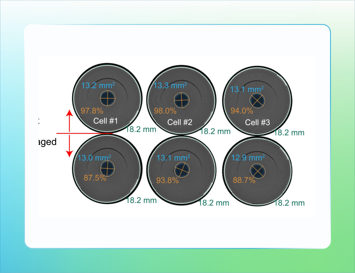
We set up a simple study: we created three groups of cylindrical 18650 cells and exposed them to either 2 minutes or 60 minutes of X-ray imaging, representing Glimpse’s typical and industry-standard CT scan durations, alongside a control group with no X-ray exposure. We then cycled all groups and looked hard for differences in performance and lifetime. We found none: capacity, resistance, and cycle life were statistically identical across all three groups. We also estimate the X-ray dose that each of these cells received and find that these doses are orders of magnitude smaller than prior literature studying beam damage from synchrotron X-ray sources.
CT scanning is a safe and nondestructive method to characterize battery cells, and Glimpse enables the throughput and image quality needed to make this technique practical at scale. Contact us to leverage high-throughput CT scanning for your own cell characterization and quality control needs.