And don’t worry about managing terabytes of scan data — we handle that for you.

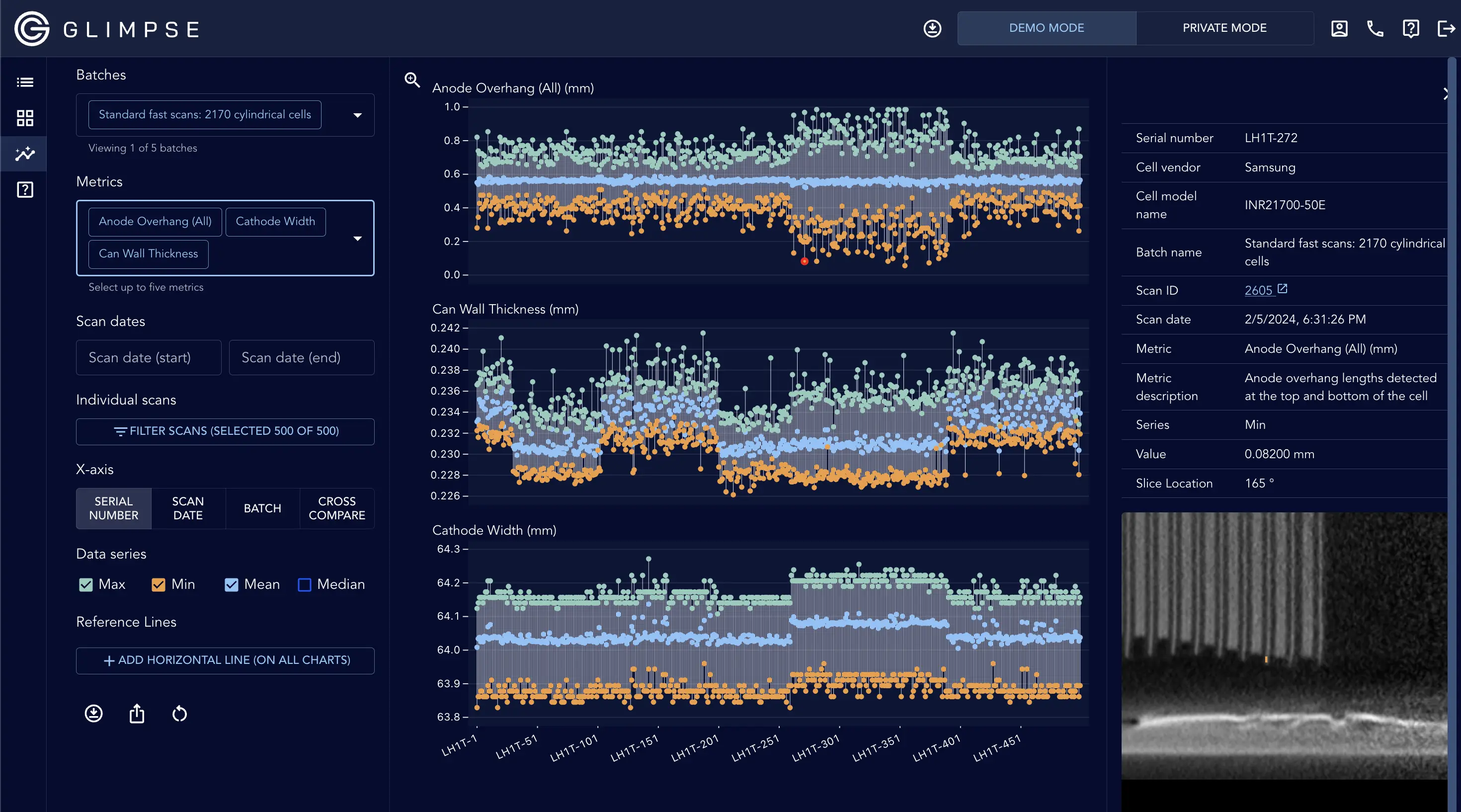
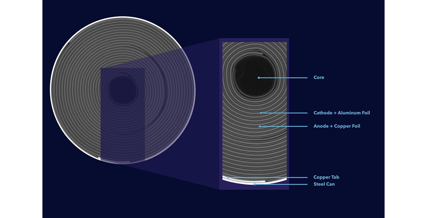
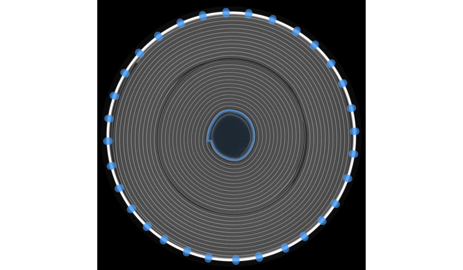
Our team has developed a full suite of generalized cell inspection algorithms by collecting millions of cross sections from the world's most diverse dataset of cell designs.

Send it to the applications team. The engineers who run the scans write these answers.
Our technology is designed to enable faster CT scanning by improving image quality through advanced image enhancement algorithms. We use trusted, conventional image processing methods that preserve the fidelity of the original data. We intentionally do not use AI or machine learning for image enhancement to avoid any risk of image hallucination or distortion. Please note that our image enhancement algorithms can be disabled upon request.

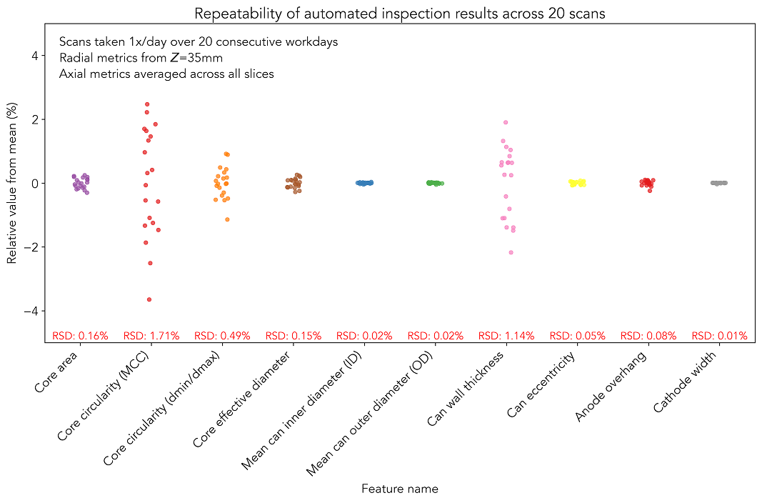
We take pride in ensuring that our automated inspection results are both accurate and precise. For instance, the below plot quantifies the repeatability of some of our standard metrics across 20 scans of the same battery cell, taken across 20 consecutive workdays:

In general, features that are only a few voxels in length will have worse repeatability than features that are tens or hundreds of voxels in length.
We evaluate algorithm repeatability on our own scanner by scanning a reference sample once per week and ensuring the results are consistent over time. We also routinely scan calibration phantoms to evaluate both dimensional accuracy and unsharpness.
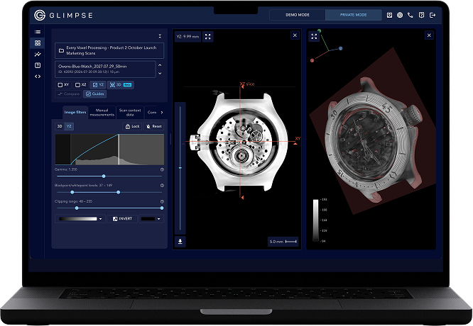
That said, no algorithm is perfect, and our algorithms will occasionally make mistakes. That’s why we’ve built the Glimpse Portal in a way that allows you to check the results of any algorithm by visualizing feature overlays over the original CT images.

Note that absolute dimensional measurements are often accurate but not formally guaranteed. Instead, our technology is primarily designed to quantify build quality and variability, detecting deviations between cells or lots with high sensitivity.
We use a Nikon XT H 225 ST 2x scanner in our lab. This scanner is an excellent choice for high-throughput CT scanning of full battery cells, largely due to Nikon’s innovative X-ray source design (read more here) and helical scanning capabilities. However, this system has been discontinued by Nikon; the new version of this system is the VOXLS 20.
If you’re planning to buy a CT scanner, please talk to us! CT scanners are complicated and expensive, and we’ve spoken to many customers who purchased a scanner that doesn’t meet their needs. We are happy to share our comprehensive guide on CT hardware selection, free of charge, even if you never use Glimpse’s products.
The absolute lower limit for voxel size for our CT scanner is about 5 µm due to the minimum spot size of our X-ray source. However, we can only achieve this voxel size for very small samples.It depends. CT cannot detect electrolyte within the jellyroll, but excess electrolyte is sometimes visible in the cell.
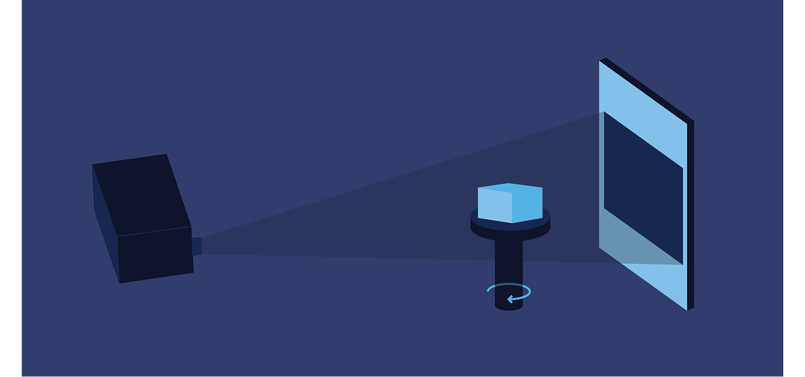
Specifically, the middle dimension of the sample determines the minimum achievable voxel size. Imagine you have an object with three dimensions of different lengths. Typically, you would place the object such that the smallest and second dimensions spin on the stage. The second dimension sets the shadow on the detector and thus sets the voxel size. Thus, we can achieve smaller voxel sizes with smaller samples.

Please see our “What drives voxel size and resolution? Can slower scans unlock improved voxel size/resolution?” FAQ question for more information.
In principle, we can scan any sample in a few seconds — but not at high image quality. Glimpse’s scan recipes attempt to optimally balance scan time and image quality. For instance, our standard cylindrical cell scan recipes take about two minutes and achieve high image quality. Please talk to us so we can help you find the best scan time/image quality tradeoff for your application (learn more here). In conventional lithium-ion batteries, both the anode (primarily graphite) and the separator (primarily polyethylene/polypropylene) are porous carbon materials. Thus, since both components have similar radiodensity and are typically wound next to each other in a jellyroll, we’re often unable to distinguish these two components in the jellyroll via CT.
We’re also working hard on improving scan time while maintaining high image quality by partnering with leading CT system integrators. For instance, the “superscanner” enables nearly 1-second scans of full cylindrical battery cells with high image quality.
We scan all major cell form factors: coin, cylindrical, pouch, and prismatic. Very large cells, such as long blade-format prismatic cells, may require multiple scans to capture in full. Contact our operations team to discuss size limits for your cells.
In general, we recommend full-cell scanning since defects can occur anywhere in a cell. That said, we support region-of-interest scanning. For instance, check out the “Ultra-high-resolution” scans visible in demo mode or view this blog post. Please talk to us to discuss further.
Generally, the optimal quantity of cells to send for scanning depends on your goals:
Glimpse’s Scan on Demand service has no minimum or maximum shipment size, so you can send as few or as many cells as you would like. If you’re interested in getting started with CT scanning through our Scan on Demand service but are uncertain about what to send us, we’re happy to talk through your goals and needs to help identify an effective scanning matrix (contact form).
Please read our blog post to learn more about shipping battery cells.
Glimpse is SOC 2 Type II certified. Our security statement can be found here, and our Vanta Trust Center page can be found here.
We prefer ACH and wire transfers, but we also accept credit cards. We will invoice you for payment once we have scanned all the cells you sent us.
We can dispose of your cells after scanning. Contact us to learn more about our cell disposal fees.
Send it to the applications team. The engineers who run the scans write these answers.

Learn from our team of battery quality experts what CT can and cannot reveal in your cells, and why.
Our published research on how cells fail and which defects matter.
Tell us the form factor, number of cells, and the questions you are trying to answer. We’ll give you a quote and scan your cells in no time.