Contact Us
Service
Service

Scan On Demand

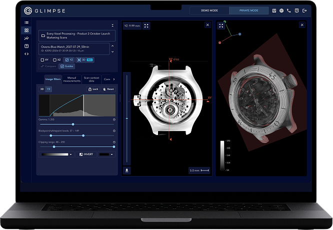
Ship us a part. We CT scan it and hand back the results in your browser — no scanner, no capital expense.
HOW IT WORKS
01
You ship it
Get quote, put your part in the mail
02
We scan it
We produce high-resolution CT scans
03
Review, measure, share
All in the Glimpse Portal
Get a scan quote
Tell us what you need scanned and we will come back with a price.
Talk to an engineer
Contact Us

CT for battery cells

Learn what CT can reveal in battery cells

Electrodes, separator, electrolyte, can, and more - learn from our team of battery quality experts what CT can and cannot reveal in your cells, and why.

What are the different components of a battery cell visible in a CT scan?
What should I be looking for in a CT scan of a battery cell?
What battery defects can CT reveal?
Can we see electrolyte via CT?
Can we see the separator via CT?
Can we see metallic particle contaminants via CT?
Can we see lithium metal / lithium plating via CT?
Do X-rays damage batteries?
Does CT work for sodium-ion, solid-state, or other next-generation battery designs?
Can I use CT to study cell mechanics as a function of state-of-charge (SOC)?
Can I use CT to study cell mechanics as a function of cycle number and/or state-of-health (SOH)?

Can’t find your question?

Send it to our engineering team and we’ll get you an answer.

Ask a question
What are the different components of a battery cell visible in a CT scan?

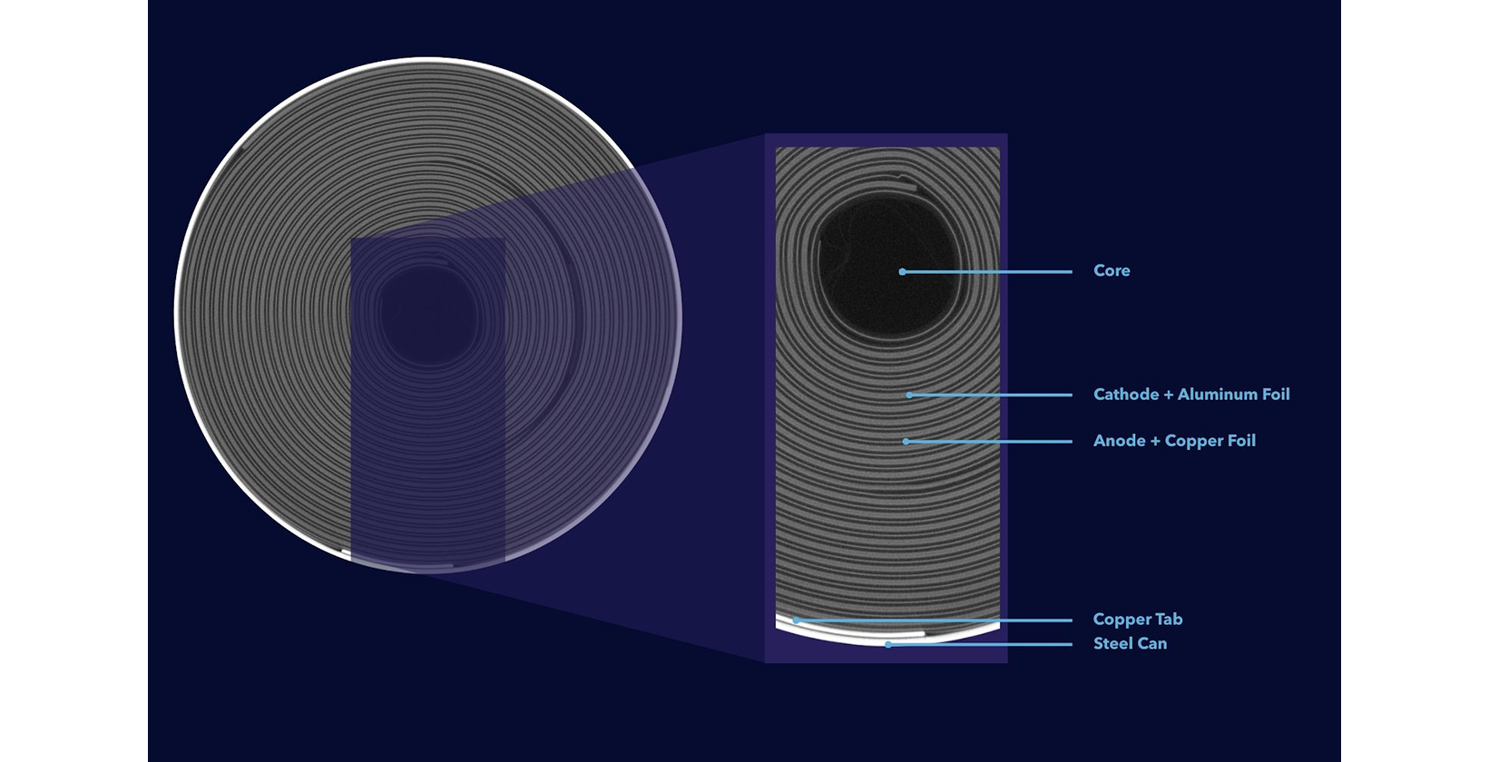
Battery cells are excellent samples for CT imaging because their key components have different elemental compositions. The brightest components in a typical cylindrical lithium-ion cell are the stainless steel can (Z = 26) and the copper tabs (Z = 29); the copper foil is also visible in high-resolution scans. The cathode is typically brighter than the anode and separator, since the cathode contains nickel or iron while the anode and separator are primarily carbon. Glimpse’s scans are optimized to distinguish these components while maintaining rapid scan times.

Diagram of a cylindrical battery cell's cross-section, labeling the core, cathode, anode, copper tab, and steel can layers.
What should I be looking for in a CT scan of a battery cell?

CT can reveal a wealth of information about a cell, including information you may not have been initially expecting.
‍
The most common use case for cell CT scanning is to check for defects — especially latent defects. Some defects that are often detected via CT include insufficient anode overhang, metallic particle contamination, core buckling, tab tears, and more (read more here).
‍
Cell CT scans can also be used to check the dimensional conformance of various cell components, including the electrodes, foils (sometimes), tabs, and can. You can see how CT can unlock this capability by checking out our automated inspection dashboards here.

What battery defects can CT reveal?

CT reveals a host of reliability- and safety-critical geometric defects, such as insufficient anode overhang, metallic particle contamination, core buckling, tab tears, and more. These defects will likely have minimal impact on performance degradation (i.e., energy or capacity fade) but can cause a battery reliability or safety event, depending on the use case. See the below image (taken from our Nature Communications publication on battery quality) to better understand the distinction between performance degradation, functional failure, and safety events:
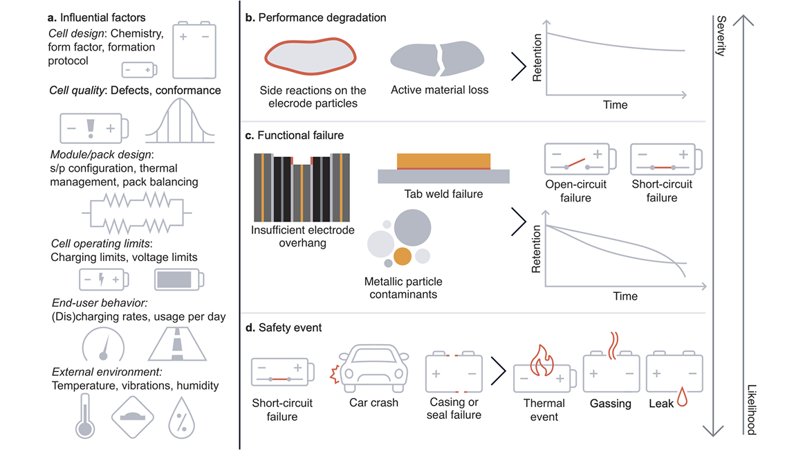
These defects are often latent, meaning they do not have an electrochemical signature until the defect has developed into a failure (e.g., an internal short circuit; read more here). In this blog post, we profile some defects that we observed in commercially-available cells.

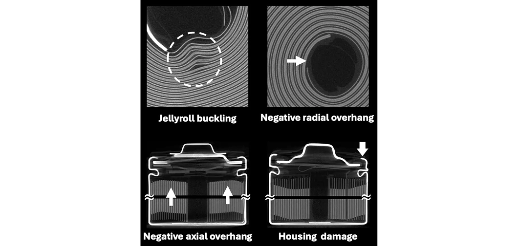
Most importantly, “critical” defects such as core buckling (“core collapse”), overhang violations, and poor crimps are major risk factors for battery reliability and/or safety events:
Additionally, “marginal” defects are possible risk factors for battery reliability and/or safety events:
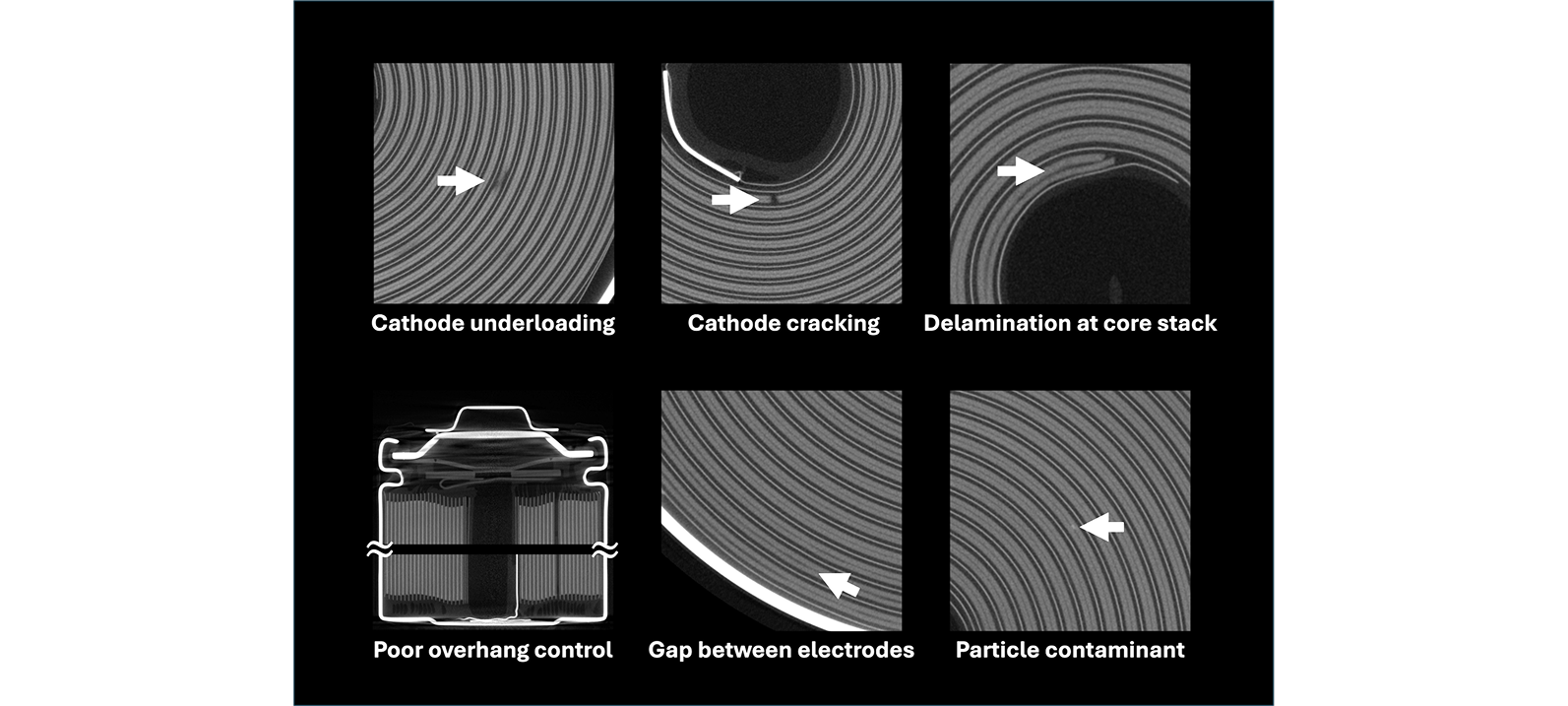
These latent defects do not have any electrochemical signature until failure (e.g., a short circuit) is imminent. We discuss the significance and safety risks of unidentified latent defects in this blog post and our Joule publication on the limits of electrochemistry for battery defect detection.
‍
The risk of an observed defect depends on many factors, such as cell chemistry, cell design, use case, and environmental conditions. More aggressive use cases necessitate more stringent defect tolerances.

Can we see electrolyte via CT?

It depends. CT cannot detect electrolyte within the jellyroll, but excess electrolyte is sometimes visible in the cell.
‍
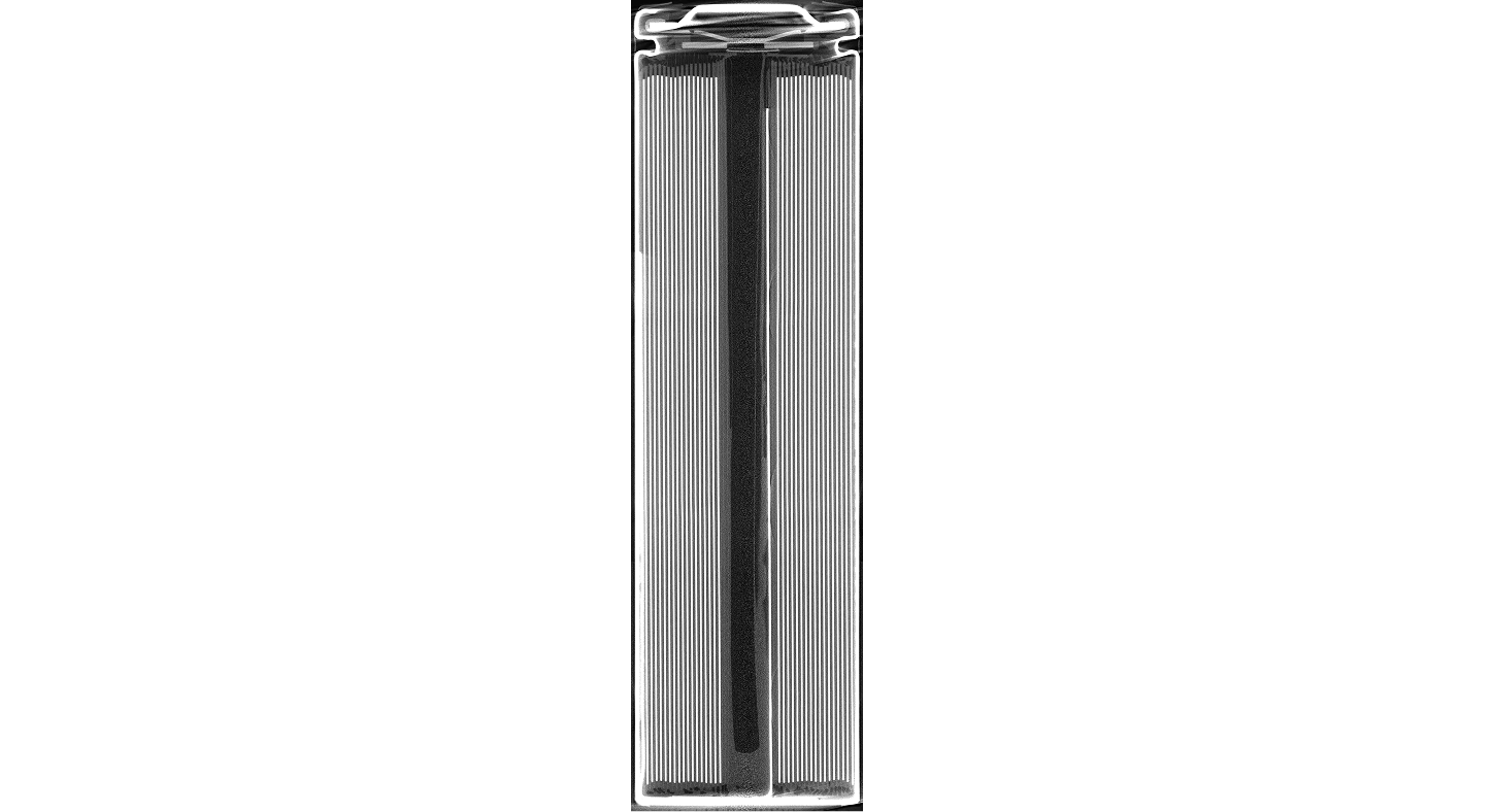
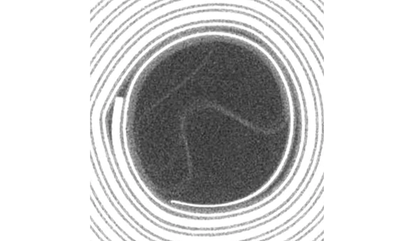
For instance, in this fresh cell from our public demo, the meniscus of the electrolyte, as well as the electrolyte soaking the separator in the core, is clearly visible (brightness enhanced for improved electrolyte visibility):

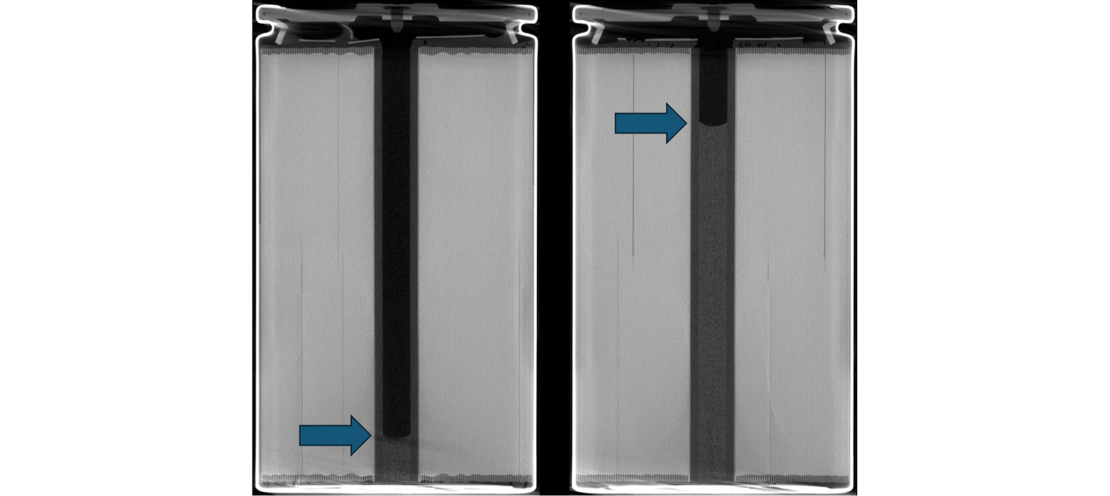
In these scans of two fresh BYD 4680 cells (brightness enhanced for improved visibility), we can clearly see the electrolyte meniscus:

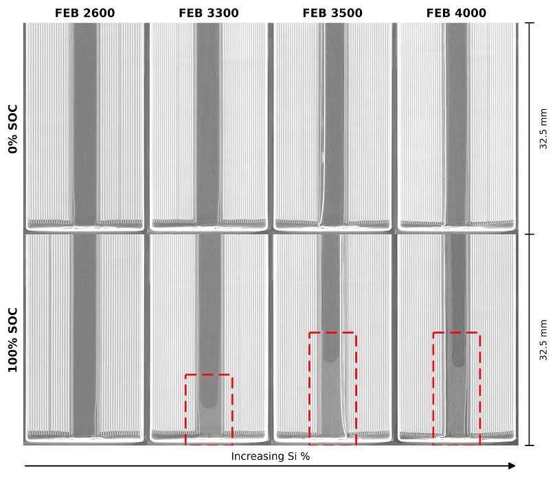
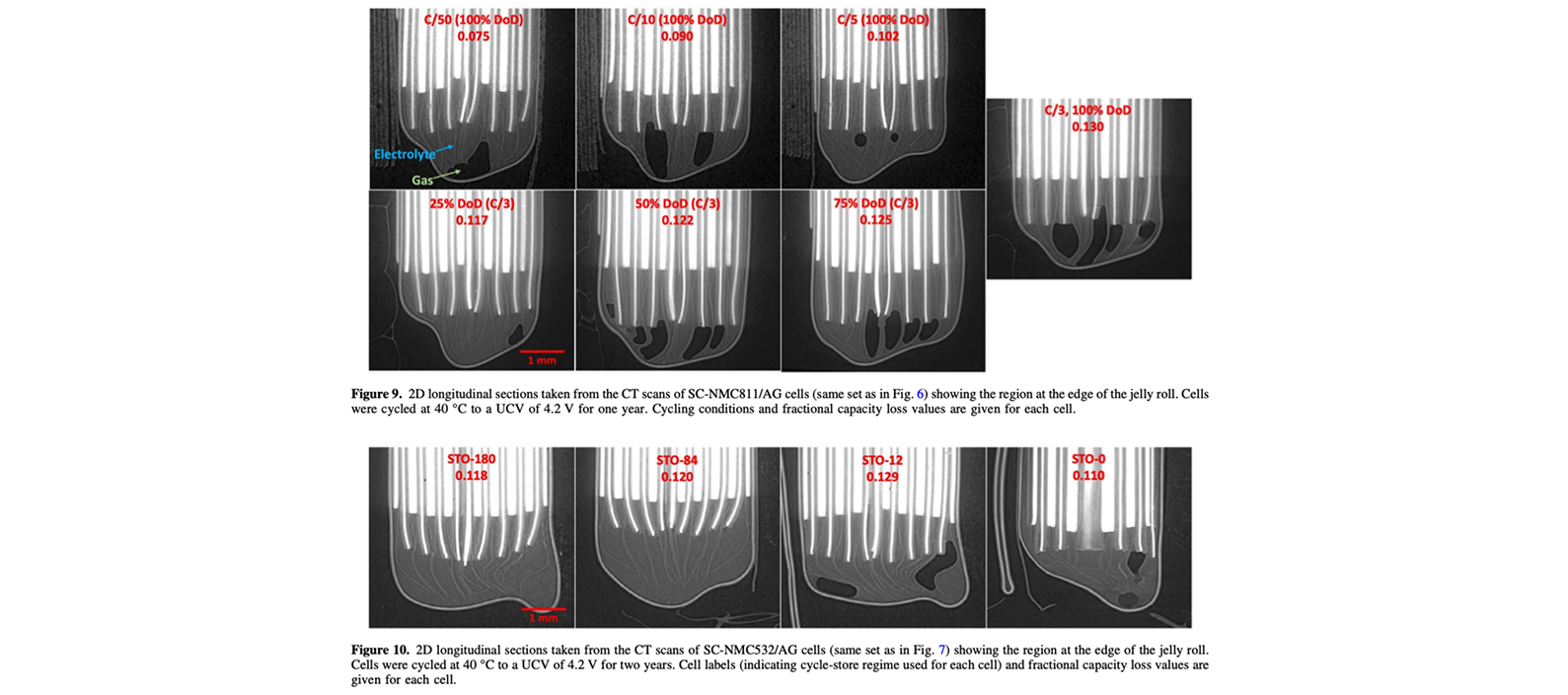
This Glimpse co-authored paper, in collaboration with the Dahn lab, illustrates how excess electrolyte in the core is often detectable via CT. The electrolyte is squeezed out of the jellyroll and into the core at high state-of-charge and in cells with high swelling:

This paper, also from the Dahn lab, uses synchrotron CT to visualize excess electrolyte in small pouch cells:

That said, detecting electrolyte via CT can be challenging. We suggest three approaches to increase electrolyte visibility:

  1. Improve contrast during scan acquisition. The two main levers for improved contrast during acquisition are (a) reducing beam voltage (see Figure 1 here) and (b) modifying source/detector settings, namely source voltage/current and detector gain, to improve dynamic range. However, lower beam voltage will reduce signal-to-noise when scanning large and/or dense cells.
  2. Charge the cell to a high state of charge (SOC). As discussed above, excess electrolyte is often visible at high SOC, especially for cells with high swelling, since the electrolyte is squeezed out of the jellyroll and into the core.
  3. Add a contrast agent to the electrolyte. Contrast agents are often used in medical imaging. For instance, this paper studied electrolyte wetting via CT on cells with a contrast agent (LiI) added to the electrolyte. LiAsF₆ is another suitable contrast agent (reference). Note that this technique is destructive (you probably won’t want to cycle this cell afterwards) and won’t work unless you’re able to fill the cells yourself, but it’s a viable approach for customers interested in a detailed understanding of their filling and soaking processes.
Can we see the separator via CT?

In conventional lithium-ion batteries, both the anode (primarily graphite) and the separator (primarily polyethylene/polypropylene) are porous carbon materials. Thus, since both components have similar radiodensity and are typically wound next to each other in a jellyroll, we’re often unable to distinguish these two components in the jellyroll via CT.

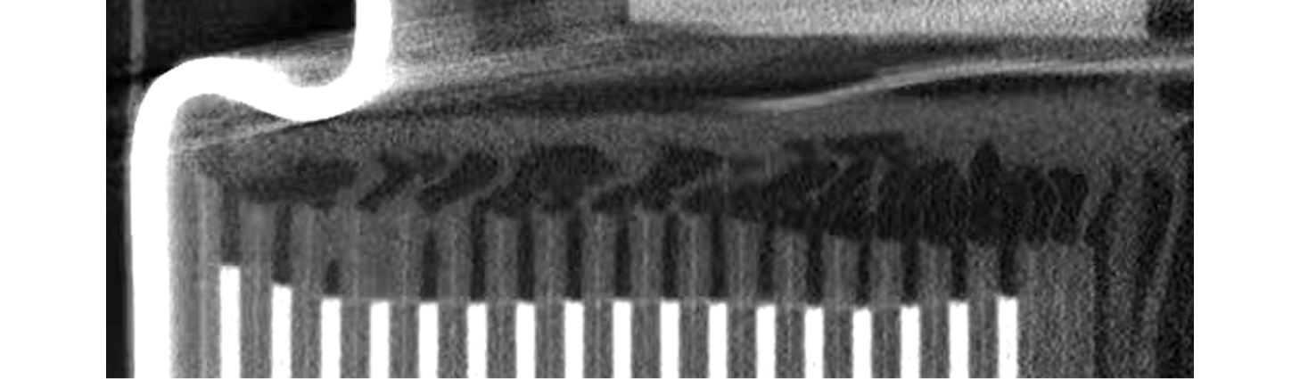
Fortunately, we can often see separator where it protrudes from the jellyroll — especially when the separator is soaked with electrolyte. Adjusting the brightness and/or contrast when viewing the scan slices may also help.

For example, the separator is visible in the core in this cylindrical cell scan:

Also, the separator is visible above the jellyroll in this cylindrical cell scan:

Can we see metallic particle contaminants via CT?

It depends. Here’s an example of a suspected particle contaminant visible in a cylindrical cell scanned via our 2-minute scanning recipe:

As you can imagine, metallic particles can be difficult to detect, especially in rapid (higher-noise) scans. The particle size, chemical composition, cell size, cell chemistry, and CT scan recipe will all impact the detectability of a metallic particle contaminant in a CT scan. Of course, larger particles will be easier to detect than smaller particles, and higher-Z contaminants (e.g., steel, copper) will be easier to detect than lower-Z contaminants (e.g., aluminum). A good rule of thumb is that particles can be reliably visualized if they are at least 3-5x the voxel size (reference).

Particle size quantification is especially challenging due to two factors:

  1. The small feature size relative to typical achievable spatial resolutions for cell CT scanning. Typically, feature sizes 5-20x larger than the voxel size are required for quantification (reference), meaning voxel sizes of 2-10 µm are required to accurately quantify the size of a 50 µm particle. See our FAQ question entitled “What’s the difference between voxel size and resolution?” for more information.
  2. “Blooming artifacts” can cause particles to appear larger than their true size.

Metallic particle contaminants are always challenging to detect. Fortunately, Glimpse’s rapid, full-cell scans offer industry-leading resolution and thus your best chance of detecting metallic particle contaminants.

Can we see lithium metal/lithium plating via CT?

Unfortunately, since the contrast in CT comes from radiodensity and lithium has very low radiodensity, lithium metal is difficult to detect via CT.
‍
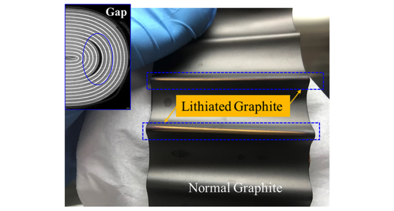
That said, large gaps between electrode layers are often a root cause of lithium plating, and these gaps are often visible via CT. For instance, this paper found that CT-detectable gaps lead to heterogeneous lithium distributions, which in turn lead to lithium plating:

This paper from Exponent also discusses how CT-detectable gaps between electrode layers often cause lithium plating.

Do X-rays damage batteries?

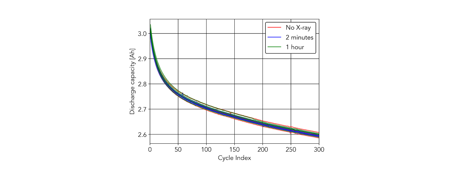
Given the number of customer questions on this topic, we decided to answer this question ourselves. We compared a control group to cells exposed to 2-minute scans as well as 60-minute scans. Try as we might, we could not identify any differences between the experimental groups. You can read the gory details here.

Does CT work for sodium-ion, solid-state, or other next-generation battery designs?

Yes. For instance, we scanned some sodium-ion cells in our public 1000-cell demo. You can see a sample scan here. We have scanned a variety of lithium-metal, solid-state, and other next-generation cells.

Broadly speaking, we can scan novel battery chemistries and designs as long as the negative and positive electrode materials have different elemental compositions — which is nearly true by definition in a battery. That said, acquiring high-quality scans of cell designs containing very high-Z elements (e.g., lanthanum) can be challenging since high-Z materials are highly attenuating and tend to introduce artifacts.

Can I use CT to study cell mechanics as a function of state-of-charge (SOC)?

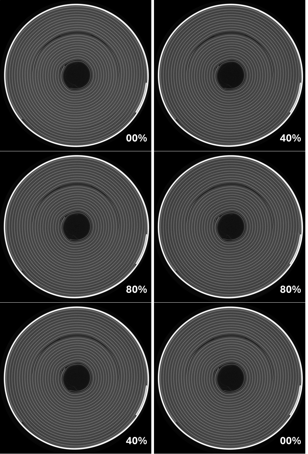
Yes, we routinely work with customers to CT scan cells as a function of SOC. For instance, see the below animation illustrating the “breathing” of a cell during a charge/discharge experiment:

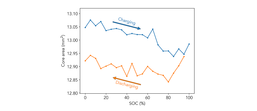
Subtle geometrical changes, such as changes in jellyroll core area, can be tracked as a function of SOC via Glimpse’s automated inspection algorithms:

Check out our paper to learn more. Glimpse performs SOC experiments in-house using our own cell cycling channels.

Can I use CT to study cell mechanics as a function of cycle number and/or state-of-health (SOH)?

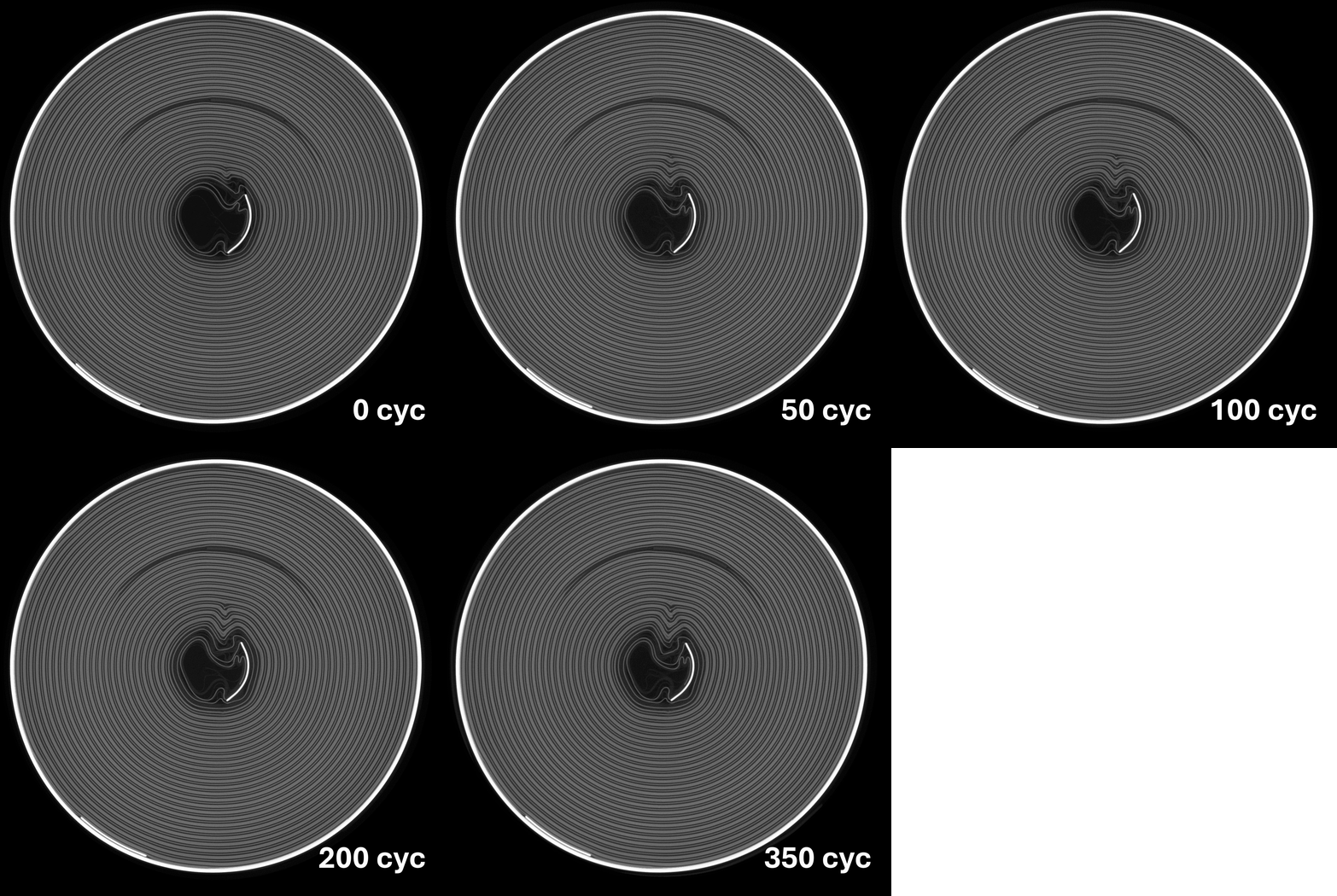
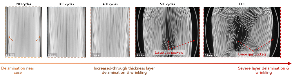
Yes, we routinely work with customers to CT scan cells as a function of cycle number and/or state of health. Check out the below animation illustrating deformation of a jellyroll over its lifetime:

We also published a paper showing the progression of jellyroll deformation in a prismatic cell over the course of a cycling experiment:

We partner with another company in our facility, Daniel Consulting Group, for cell cycling experiments. In general, we highly recommend CT scanning cells before starting a cycling test. Read more in our blog post on this topic.

Can’t find your question?

Send it to our engineering team and we’ll get you an answer.

Ask a question

Keep Learning

Dive deeper on battery quality, CT,
and how our customers use the Glimpse Portal

CT theory

What CT can reveal inside a part and why.

Learn more

Battery quality science corner

Our published research on how cells fail and which defects matter.

Learn more

Case studies

Learn how our customers are using Glimpse to unlock battery quality at scale.

Learn more

Ready when you are

Start scanning your cells now.

Get the Glimpse Portal Battery package for your CT system or send your cells to one of our Scan On Demand centers.

Not sure where to start? Talk to us.