At Glimpse, we pride ourselves in providing industry-leading scan time and image quality. But what do we mean by “image quality”? In reality, image quality in CT is a complex, messy, multifaceted topic, and no single metric can be used to encapsulate image quality. In this post, we’ll discuss and illustrate various aspects of CT image quality and the tradeoffs inherent to this technique.
Perhaps the most important thing to know about image quality in X-ray imaging and CT is that it’s highly task-dependent and, for better or worse, identifying task-relevant image quality requires human expertise. Jim, one of Glimpse’s founding engineers, has spent most of his career in medical imaging and most recently served as the Director of AI & Computer Vision at a large medical imaging company. He is a true X-ray imaging and computer vision expert. In his words: “There are 1000-page textbooks on image quality for medical imaging, but in practice you should toss them aside and just evaluate if the image quality is suitable for the task at hand.”
Here, we’ll discuss four image quality metrics: voxel size, signal-to-noise ratio, sharpness, and artifacts. These four metrics are four of many, many image quality indicators, but they are perhaps the four of greatest interest to the battery cell CT scanning use case.
The sample images below are taken from a cell in our free demo of the Glimpse Portal.
Voxel size
A voxel (“volume pixel”) is a 3D version of a pixel. Since voxels are nearly always cubes (as opposed to rectangular prisms), the “voxel size” is typically defined by the length of one side of the cube (e.g., “20 µm”). Just like how each pixel contains one color, each voxel contains one grayscale value. See below for images with small and large voxel size.
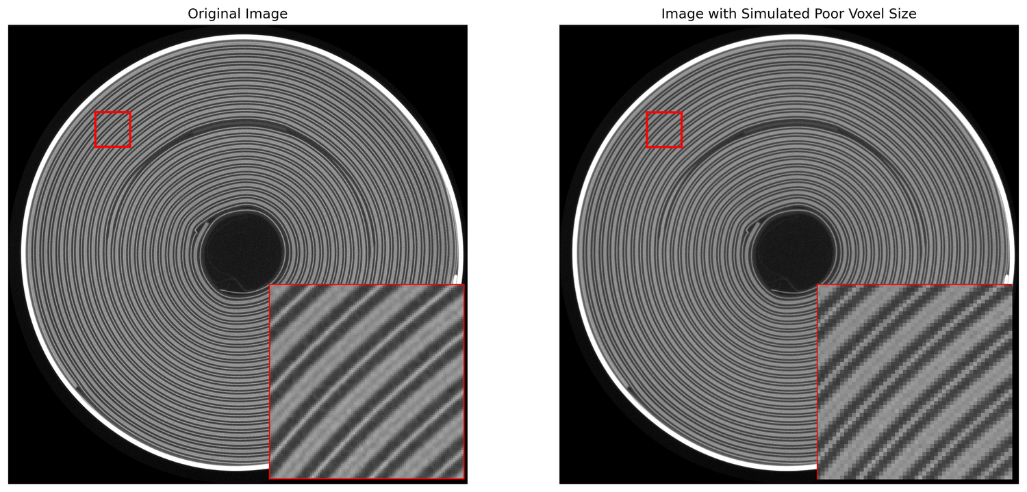
Importantly, voxel size is not the same as resolution! These metrics are often conflated. Voxel size defines the physical dimensions of each voxel, while resolution reflects the ability to distinguish fine details in an image. Though smaller voxel sizes often suggest higher resolution, factors like sharpness, noise, and artifacts can play a significant role in determining the resolution. Read here and here to learn more.
Note that small voxel sizes result in large file sizes since the sample is discretized over smaller intervals.
Signal-to-noise ratio (SNR)
The signal-to-noise ratio, or SNR, reflects the strength of the signal relative to the noise. Low SNR makes segmentation and feature extraction difficult. Read more about SNR here, and see below for images with high and low SNR.

Sharpness
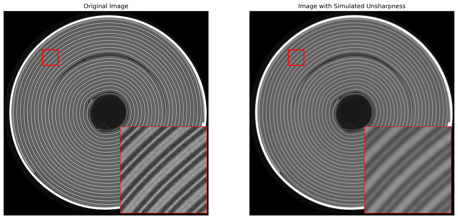
Sharpness refers to the clarity and detail of the edges within an image. In other words, sharpness describes the ability to resolve fine structures in an image, particularly where the density abruptly changes (i.e., at the boundary between two materials). Images with poor sharpness will have blurred edges and thus are difficult to extract features from. Read more about resolution and sharpness here, and see below for images with high and low sharpness.
Artifacts
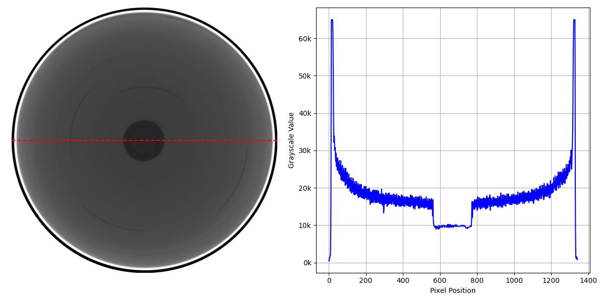
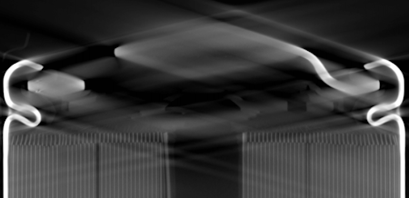
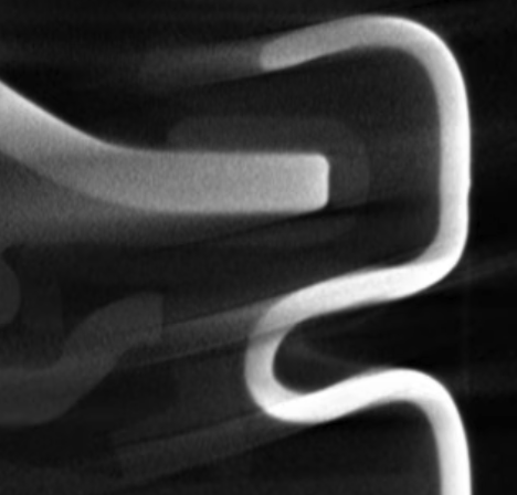
CT reconstruction is an “inverse problem”, meaning its goal is to reconstruct the ground truth of an object from observations resulting from that ground truth (e.g., 2D projection X-ray images). Algorithms to solve inverse problems, such as CT reconstruction, must make assumptions about the physical world that do not always hold perfectly. As a result, “artifacts”, or nonphysical structures in the reconstruction, can occur and convolute scan interpretation and automated scan analysis. Many artifacts can be controlled/reduced/eliminated, but some are unavoidable. See here and here to learn more about common CT artifacts. We illustrate a few of the most common artifacts in the images below:
Tradeoffs in image quality
Battery-related press releases are infamous for exaggerating one parameter (e.g., fast charging capability) at the expense of others (e.g., energy, lifetime). Similarly, a CT scan might have impressive voxel size but poor SNR and sharpness. Optimizing a CT scan to have high overall image quality requires a deep understanding of both the fundamentals of CT imaging as well as the imaging task at hand (e.g., detecting subtle metallic particle contaminants).
Of course, a critical parameter to consider when optimizing image quality is scan time. Naturally, slower scans can yield better image quality. This tradeoff can be visualized via the below images. Here, we scanned the same cell with four different scan recipes, with scan times ranging from 1 minute to 16 minutes. While the overall structure of the cell can be readily observed in all scans, the slower scans are clearly less noisy. Furthermore, two subtle features are much clearer in the slower scans than the faster scans: first, the separator is faint but visible in the slow scans but not visible in the fast scans, and second, the structure of the chipped cathode is much clearer in the 16-minute scan. That said, the overall structure of the battery is very clear even after the 1-minute scan.
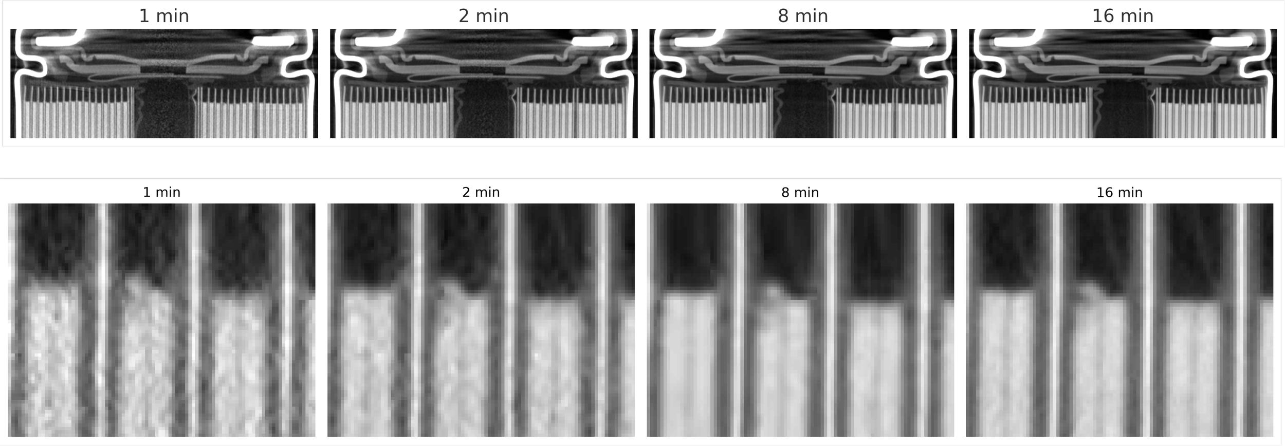
We generally recommend approaching this scan time–image quality tradeoff by constraining one variable and then optimizing the other— e.g., setting a scan time based on a desired throughput level or setting a required image quality level based on specific defects of interest.
A final parameter to consider is the region of interest—e.g., is the goal to scan the entire cell or just, say, the overhang region? Sometimes, a region-of-interest scan is suitable, but we generally recommend full-cell scanning since defects can occur nearly anywhere in a cell. Often, marketing materials showcasing impressive CT images are in fact only scanning a small portion of the cell.
Summary
As we’ve discussed, optimizing image quality is a complex, multidimensional problem. In many ways, CT imaging is like photography in that the right hardware is necessary, but not sufficient, for obtaining high-quality images; expertise in acquiring and processing images is also essential.
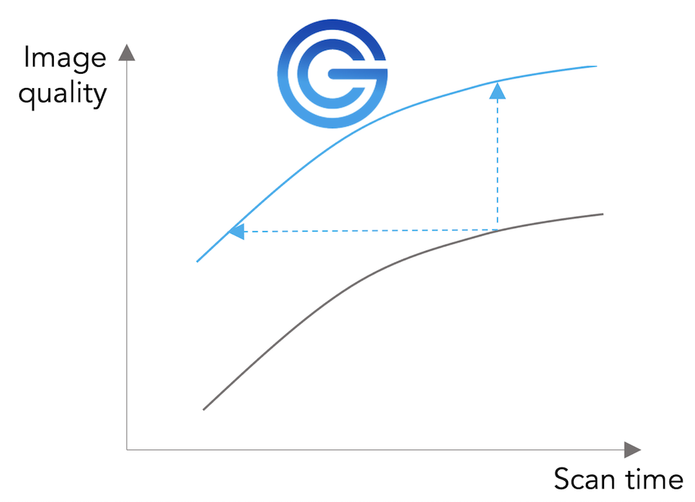
At Glimpse, one of our primary technical goals is to improve the Pareto front of scan time and image quality for our customers (and we’re actively working on improving this Pareto front even further!).
Again, because image quality is multifaceted, task-specific, and can’t be encapsulated by a single metric, Glimpse applies its battery expertise to help make these image quality tradeoffs with our customers. Contact us if you’d like to learn more about how Glimpse can help you optimize scan time and image quality for your application.


